Airway-focused orthodontics is gaining traction as a proactive approach to diagnosing and treating malocclusions with a greater emphasis on airway health. The integration of cone beam computed tomography (CBCT) and sleep studies has revolutionized early orthodontic intervention, allowing practitioners to assess airway obstructions, detect sleep-disordered breathing (SDB), and implement treatment strategies that promote optimal growth and development. This article explores the role of CBCT and sleep studies in early orthodontic intervention and their impact on long-term health outcomes.
Understanding Airway-Focused Orthodontics
Traditional orthodontics primarily focuses on teeth alignment and bite correction. However, airway-focused orthodontics extends beyond esthetics and function to address breathing-related concerns, such as obstructive sleep apnea (OSA) and other forms of SDB. Research indicates that narrow airways, mouth breathing, and poor tongue posture can contribute to malocclusions, skeletal deficiencies, and overall systemic health issues (Guilleminault et al., 2016).
The Role of CBCT in Early Orthodontic Diagnosis
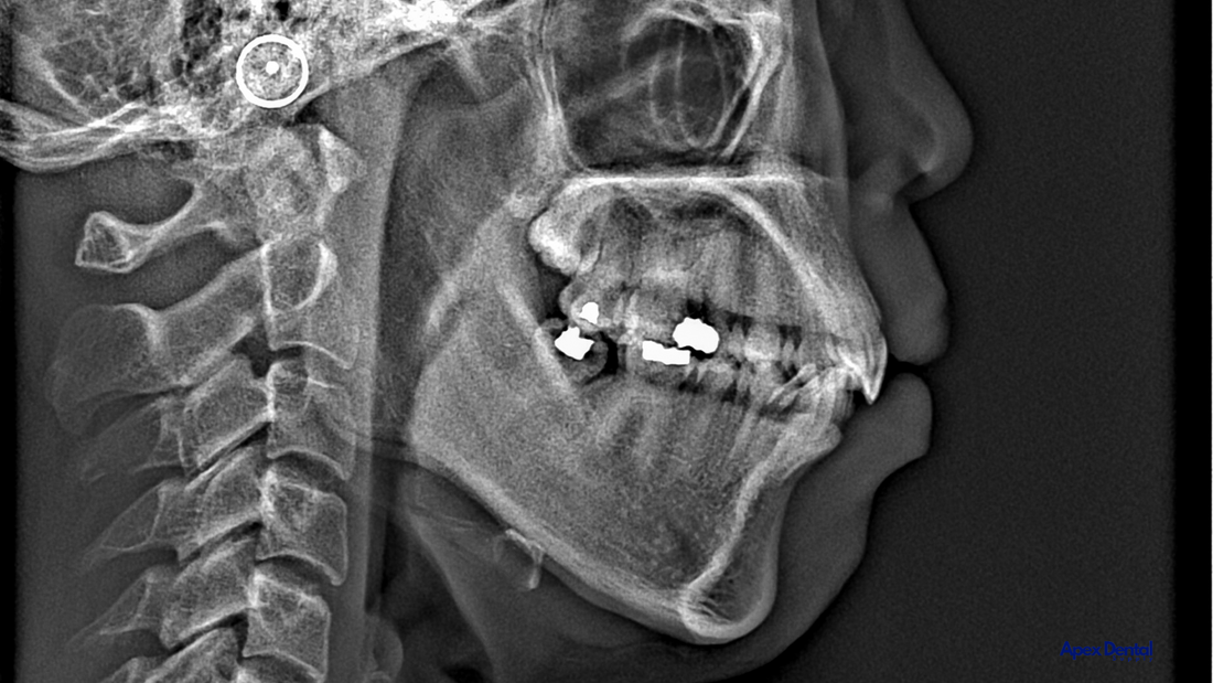
CBCT has revolutionized orthodontic imaging by providing three-dimensional (3D) visualization of craniofacial structures. Unlike conventional two-dimensional radiographs, CBCT allows practitioners to assess:
-
Airway volume and dimensions: Identifying airway constrictions or obstructions.
-
Skeletal morphology: Evaluating maxillary and mandibular development.
-
Nasal and sinus abnormalities: Detecting deviations or blockages that may affect breathing.
-
Tongue space and posture: Understanding tongue placement and its impact on airway patency.
Several studies underscore the significance of CBCT in diagnosing airway issues. A study by El and Palomo (2010) demonstrated a correlation between reduced airway volume and Class II skeletal patterns, highlighting the importance of early intervention.
Sleep Studies: Diagnosing Sleep-Disordered Breathing
Polysomnography (PSG) is the gold standard for diagnosing sleep disorders, including OSA. However, home sleep apnea tests (HSATs) are increasingly being utilized for children and adolescents. Sleep studies provide vital information such as:
-
Apnea-hypopnea index (AHI): Measuring the severity of airway obstruction.
-
Oxygen saturation levels: Assessing the impact of airway restrictions on oxygen intake.
-
Sleep fragmentation: Evaluating disruptions in sleep architecture due to breathing difficulties.
A 2020 study by Lee et al. found that children with enlarged adenoids and tonsils, coupled with narrow maxillary arches, exhibited higher AHI scores, reinforcing the need for orthodontic evaluation in pediatric patients with suspected SDB.
Early Orthodontic Intervention for Airway Optimization
By leveraging CBCT and sleep study data, orthodontists can implement growth modification techniques to improve airway function. These interventions include:
-
Maxillary expansion: Palatal expansion increases nasal airway volume, reducing resistance to airflow (Chang et al., 2013).
-
Mandibular advancement appliances (MAAs): Often used in growing patients to encourage proper jaw positioning.
-
Myofunctional therapy: Exercises aimed at strengthening orofacial muscles to promote nasal breathing and correct tongue posture.
Long-Term Health Benefits
Early identification and treatment of airway issues can prevent chronic health complications, including:
-
Cognitive impairments: Studies show that untreated SDB in children can lead to attention deficits and learning difficulties (Kushida et al., 2012).
-
Cardiovascular risks: Persistent airway obstructions can contribute to hypertension and cardiovascular disease later in life.
-
Craniofacial growth deficiencies: Addressing airway problems early fosters proper skeletal development and reduces the likelihood of severe malocclusions in adulthood.

The integration of CBCT imaging and sleep studies has significantly enhanced early orthodontic diagnosis and treatment planning. Airway-focused orthodontics emphasizes functional breathing and skeletal harmony, reducing the risk of long-term health complications. By identifying and addressing airway obstructions in children, orthodontists can contribute to better sleep quality, cognitive function, and overall well-being. As technology continues to advance, incorporating airway assessments into routine orthodontic care will become an industry standard, ultimately improving patient outcomes.
References
-
Chang, S. J., Kim, T. W., & Shin, S. J. (2013). Effects of rapid maxillary expansion on upper airway dimensions. American Journal of Orthodontics and Dentofacial Orthopedics, 143(1), 106-112.
-
El, H., & Palomo, J. M. (2010). Airway volume for different dentofacial skeletal patterns. American Journal of Orthodontics and Dentofacial Orthopedics, 137(4), 470-475.
-
Guilleminault, C., Huang, Y. S., & Monteyrol, P. J. (2016). Pediatric obstructive sleep apnea: From clinical symptomatology to treatment. Pediatric Pulmonology, 51(4), 322-331.
-
Kushida, C. A., Littner, M. R., & Morgenthaler, T. (2012). Practice parameters for the indications for polysomnography and related procedures. Sleep, 28(4), 499-521.
-
Lee, R. W. W., Sutherland, K., & Chan, A. S. L. (2020). Relationship between craniofacial structures and obstructive sleep apnea severity in children. Sleep Medicine, 75, 63-70.